

 6 July 2026
 BREAST SURGERY
 0 Comments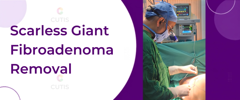
When you think of a breast tumour, or lump, the word “cancer” usually comes to mind. Given how rapidly breast cancer rates are rising, that fear is not entirely unwarranted. So imagine the fear a fast-growing breast tumour would cause in a prepubescent little girl. In this case, the tumour was so large that it was compressing the developing breast tissue, to the point where it was interfering with the normal breast development of a twelve-year-old. This case came as a surprise to us too.

A few weeks earlier, a 12-year-old girl came to our OPD with her mother. Her complaint was a large, very rapidly growing tumour in her right breast, measuring 10 x 8 cm. Surprisingly, given its size, the pain was negligible. In fact, that lack of significant pain was the reason for her late presentation to us. When her mother first noticed the tumour two months earlier, it was very small. She ignored it at first and waited nearly six weeks, hoping it would disappear on its own. But when it grew large enough to compress the developing normal breast tissue and thin the overlying skin of the areola, her mother brought her to us for treatment.
On examination, we found a firm, very large, non-tender, oval mass within the little girl's right breast. We investigated, and the radiological findings pointed to a benign, non-cancerous breast tumour. We suspected she was suffering from a giant juvenile fibroadenoma of the breast. Once we confirmed the diagnosis and reassured the patient's mother about the necessity of surgery, we decided to move forward and planned the procedure.
When removing such a tumour, the goal is to achieve the objectives of treatment in the best interest of the patient. In this case, those objectives were particularly challenging to achieve. Let's see why.
The first and most important objective was the complete removal of the tumour, without leaving any bits attached to the normal breast tissue, to prevent recurrence. The second objective seemed to contradict the first yet was just as important: to avoid injuring the normal breast tissue. Any injury to that tissue during removal could seriously affect the future development of the breast in a 12-year-old girl and lead to deformity.
The third objective may not seem relevant at such a young age, but ignoring it could leave the patient with permanent mental trauma: the scar of surgery. Let's get into the story of how we achieved all three.
There is, of course, no substitute for qualified training, surgical skill, and proper planning. But the game changer for this surgery was the use of magnification. While its role cannot be overstated, achieving all three objectives also required several things:
- proper planning and preparation for surgery
- careful selection of the site for the incision
- gentle dissection of the tissues to separate the tumour
- avoiding injury to normal breast tissue by ensuring consistent haemostasis (stopping the bleeding)
- perfect closure of the tissues
- attentive post-operative care
First, we chose to make the incision in the crease below the breast, the inframammary crease, which becomes inconspicuous over time. Normally, our incision for a breast tumour is centred at the junction of the areola, the dark skin around the nipple, and the normal skin. This is called a periareolar incision. But as noted, the tumour was larger, at 10 x 8 cm, than the breast itself, so a periareolar incision would have been too small to deliver it. And extending the incision directly over the breast tissue and the tumour would have caused undesirable scarring and disfigurement. So we chose the most challenging, yet nearly scarless and safe, approach: from beneath the breast, in its natural crease.
In terms of scarring and patient satisfaction, this was without doubt the best approach for removing the tumour. But it was technically very challenging for the surgeon to reach such a large breast tumour from below and to do so using the 4X magnification needed to prevent injury to the normal breast tissue while ensuring the tumour was removed completely and intact.
Working with magnification in such a difficult scenario puts a tremendous load on the surgeon's neck and back muscles. He is looking from below upwards, while constantly maintaining a distance of 45 cm from the magnifying loupes to the operative field, and all within a restricted circular field of vision barely 8 cm wide. In such an extreme situation, giving up the magnifying loupes would certainly have made the whole surgery easier for the surgeon. So why insist on magnification despite the tremendous strain on the back and neck muscles in such awkward working conditions? Simply to fulfil the first two objectives of surgery: to completely remove the tumour, and to avoid injuring the normal breast tissue.

Let us see how magnification helped us achieve both of these opposing objectives. Magnification in the range of 4X gives the surgeon as good a view as an operating microscope at the same magnification. This lets the surgeon clearly identify, isolate, and control the abnormal blood vessels around the tumour before they bleed and make the dissection messy. (Abnormal growth of blood vessels is the norm in large tumours anywhere in the body.) Magnification also helps the surgeon clearly see the definite, natural plane of separation between the tumour and the normal breast tissue, which is otherwise very flimsy and nearly invisible to the naked eye. On top of that, with magnification, separating the tumour from the normal breast tissue becomes a bloodless, seamless operation with no collateral damage. These advantages are exactly why we never even consider operating without magnifying loupes, even in extreme situations. It is the magnification, along with precise surgical skill, that helps us navigate such difficult and stormy situations.

In the end, we happily completed the surgery. We removed the tumour intact, without injuring the developing normal breast tissue, and with negligible blood loss, achieving an excellent, almost invisible scar hidden in the crease below the breast. What came as a happy surprise was the speed at which the compressed normal breast tissue resumed its growth. Within just six weeks after surgery, it had caught up to a growth comparable with the normal, unaffected breast.
Seeing the patient's quality of life restored, with minimal scarring and excellent healing, brings us real satisfaction and makes us forget the challenges we faced during the surgery.
In fact, it is this feeling of pride and joy that drives a surgeon to keep pushing his boundaries and to keep doing better.

